 Your Rights
I recognize that patient has certain rights as individuals, as members of community and society, and would like to do everything possible to respect these rights within the context of receiving retina care. With Dr Pratik Mahajan every patient has a right to receive the best possible care, deleivered in a caring and compassionate manner.I always encourage patients to understand the nature of the disease and take active participaton in their disease management. I respect the following rights.
1. Privacy during consultation, examination and medical procedures
2. Confidentiality of all health information
3. To be informed about the treatment charges and expeted costs
4. Care without the discrimination of socioeconomic status, race, color,religion , gender , age, nationality or physical or mental disability.
5. Emotional and physical, protection and support for disabled, elderly or a mentally retarded child.
6. Refuse treatment
7. Obtain a disharge summary according to the hospital policy
8. Understand the possible risks , benefits or side-effects or outcomes of the treatment.
9. To understand the various treatment options in his/her own language and make informed decision in one s own benefit.
 Prepare for your Consultation
1. Its always better to take prior appointment. Please call Tina at 0777 108 9999.
2. Please come with your old health records about the eye or other systemic problems
3. Please take out time of about 2 hrs since the pupillary dialatation process may take time
4. If you are putting any anti glaucoma eye drops pease continue them as per schedule , even in the morning of consultation day, if it was scheduled. This will help assess the IOP whih is being maintained.
5. If you are diabetic , please take your breakfast and insulin /oral medications on time.Please bring your diabetes medicines with you.Please bring a small tiffin from your home , since pupillary dialatation may require longer time.
6. Its good if you bring your recent blood sugar , lipid profile or HbA1c reports with you.
7. Please bring dark goggles . Please call someone to pick you up and take you to home.
Before Your Fundus Angiography
Fundus Fluorescein Angiography is a test to study the blood vessels of the eye.This is a test to show more information about the blood circulation and condition of the back of your eyes. It helps Dr Pratik to decide the best form of treatment or management
1. Before the test, eat, drink and take all medicines as normal.However Dr Pratik advices nil- by- mouth just one hour before your scheduled appointment.
2. Bring all the medicines with you.
3. If possible, bring someone with you. If not, please bring along a telephone number of next of kin, or chosen contact person.
4. On arrival, you will be asked some questions about your general health, including any allergy. Please tell if you are allergic to specific things or your are pregnant or lactating mother.
5. The test will be fully explained before asking you to sign your consent.
6. The test may be sometimes deferred due to age, history of allergy or high serum creatinine levels or pregnancy.
7. If you are on dialysis for renal failure, we always have a word with your nephrologist and plan the test accordingly.
8. Eye drops will be put into both your eyes to dilate the pupils. They take 20 minutes to one hour to work. They will blur your vision and make light brighter for approximately 2-3 hours. Therefore, it is advisable to bring sunglasses and not to drive.
9. When your pupils are dialted, the photographer will position you comfortably sitting in front of the camera. A series of images will be taken of the back of your eyes. Most of the time an anesthetist is there with you during the test.
10. After 1st "base line"images, a small injection of fluroscein will be given into your forearm. This travel to your eyes within seconds and highlights the blood circulation during a series of images taken over the next few minutes. Then wait for 5 minutes. Some final images are taken after the circulating fluorescein has left the eyes .
11. This is is then complete and we ask you to sit within the department for 15 minutes till you become comfortable.
12. You will have a yellowish discoloration of urine/ sweat and yellowish vision. Please dont worry about that.
13. 1 in 10 patients feel nauseated or light headed for a few seconds to a few minutes shortly after the injection. The feeling usually passes quickly.
14. Some patients feel an itching or tingling sensation, or shortness of breath.
15. We provide fast and effective treatment for all side effects lasting more than a few minutes. Although extremely rare, a serious anaphylaxis reaction has been reported on 1 in 200,000 patients.
16. Please feel free to ask the analysis and interpretation of test. Please plan further treatment and make informed decision about the same. Always leave the premises after taking a follow-up date.
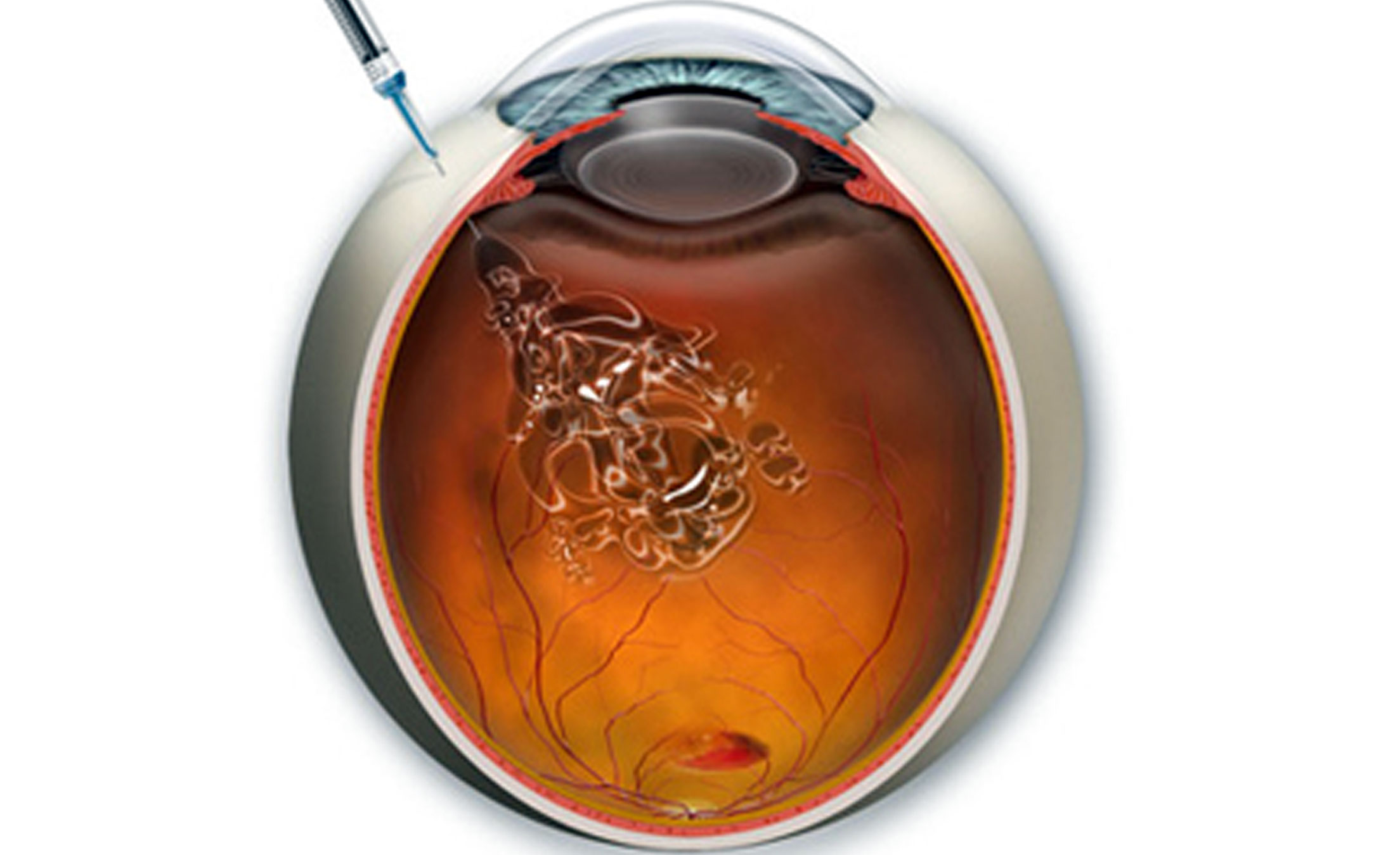 Intravitreal Injections
THE INJECTIONS FOR MACULAR DEGENERATION: Do you feel confused ?
As many of my patients who tell me ,various doctors often have very different injection protocols, making this topic very confusing for many patients. Currently, most retina surgeons adhere to one of three treatment methods:
Continued interval therapy or 'monthly' -injection given every 4 weeks (Avastin and Lucentis) or Eylea which is coming up… every 8 weeks.
'Treat and extend' -monthly injections until clinically stable for physician, then increasing intervals between injections.
'PRN' or as needed- injections given monthly until deemed clinically stable by physican and then retreat if patient becomes symptomatic or demonstrates new activity of disease.
So why the different methods?
In 2006, the results of ANCHOR and MARINA (2 large trials looking at effectiveness of Lucentis dosed monthly for AMD) left us surprised by providing visual improvements and stability never before seen. Since then, other trials have followed looking at ways to obtain equal outcomes with fewer injections, indeed.
Studies repeatedly show that manufacturer recommended monthly therapy results in the best visual outcomes but at the price of more frequent injections. In fact, patients from the original trials who were switched from monthly injections to as needed after 2 years, lost half of their visual gains in the subsequent year, conferring that the best way to maintain vision is to prevent the disease from progressing.
More recently the National Eye Institute sponsored randomized trial called the Comparison Of Age-related Macular Degeneration Treatments Trial (CATT) compared efficacy of Avastin (bevacizumab) to Lucentis (ranibizumab) dosed monthly or as needed. At two years, this study showed that when compared head to head, PRN treatment was inferior to monthly and resulted in poorer visual outcomes.
While treat and extend seems like a reasonable compromise between the two treatment regimens, unfortunately, no large clinical trials have been performed to compare the outcomes of this technique against monthly therapy. By virtue of the fact that this technique extends the interval outside the 4 week therapeutic window of the drug, patients are left unprotected with an increasing risk of recurrence and irrecoverable vision loss, and in fact, Dr Pratik has seen patients develop bleeding when patient loses follow up and presents at 5 to 8 weeks!. So while we will sometimes extend patients beyond monthly therapy if they have remained stable for several months, it is important that they understand and accept the potential risks.
OZURDEX
Ozurdex is a steroid implant which has been approved for use in retinal vein occlusions and uveitis.For more information visit www.ozurdex.com.
LUCENTIS
For more information on lucentis please visit www.lucentis.com
Before Intravitreal Injection:
Come after a shower and head and face-wash in the morning of your appointment. Have a light breakfast.
Dr Pratik always advices antibiotic eye drops to be instilled frequently in both eyes, before injection.
Use of Proparacaine 0.5% eye drops and 30 gauge needle makes injection an essentially painless procedure in the hands of
Dr. Pratik.
After Intravitreal injection:
Your eye will patched for an hour or two. Sometimes you are advised to rest in a head-elevated position for one day.
Put antibiotic eye drops and antiglaucoma eye drops as per schedule .
Don't let water go inside your eye for next 3 days.
You must follow up the next day and after one week.
Dont worry if feel floaters in your visual field.
However if you develop inceasing redness, discharge , pain or blurring of vision please contact 7771089999.
 Vitreoretina Surgery
Introduction
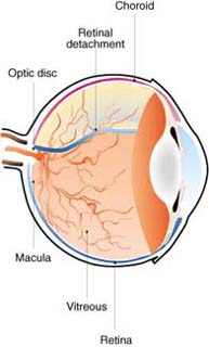
The retina lines the inside of the eye and is a thin tissue composed of layers of light-sensitive cells which send visual information to the brain. The retina is held in place by the vitreous humour which is a transparent gel composed of water and collagen and lies in the centre of the globe of the eye between the retina and lens. A retinal detachment occurs when the retina pulls away from inside wall of the eye causing loss of vision. This is often due to a hole or tear in the retina produced when the vitreous liquifies with the aging process. Trauma may also lead to retinal detachment. The vitreous may also become filled with blood, particularly in association with severe diabetic eye disease where traction may detach localised areas of retina
THE OPERATION
The indications for retinal surgery include:
1. Removal of vitreous haemorrhage,
2. Peeling of epiretinal membranes,
3. Treatment of macula holes
and most frequently retinal detachment.
Small holes or tears in the retina may be treated with laser photocoagulation or cryopexy (freezing). Laser photocoagulation consists of pinpoints of laser which creates minute burns around a small hole in order to help the retina adhere to the wall of the eye. It can also be used to treat areas of the retina which have a poor blood supply.
Cryopexy: is a procedure which freezes the area around a hole to the wall of the eye.
Scleral buckling :is a surgical procedure used in large retinal detachments in which a synthetic band is placed around the outside of the eye in order to push the wall of the eye against the detached retina.
Vitrectomy :is the surgical removal of diseased vitreous and the insertion of an artificial substance to push the retina back against the wall of the eye. The substance may consist of an expandable gas or silicone oil. The gas is slowly absorbed by the body after a couple of weeks. The silicone oil may be removed surgically when Dr Pratik decides it is necessary.
THE RISKS OF SURGERY
1. Reaction to anaesthetic agents
2. Infection
3. Bleeding
4. Cataract
5. Secondary Glaucoma
6. Failure to attain the intended outcome
7. Blindness
8. Need of repeat surgeries
SPECIFIC PRE-OPERATIVE PREPARATION
You will not need to compulsory be nil-by-mouth before the operation.Most of time Dr Pratik prefers under local anesthesia with mild sedation given by a senior anesthetist.A light breakfast can be taken in the morning if no specific instructions have been given. You should have a shower, shave and wash your hair before the operation. You will be prescribed frequent antibiotic eye drops before surgery.
LENGTH OF TIME IN HOSPITAL
From morning till evening.
AFTER THE OPERATION
Pain relief :Tablets are usually sufficient to control your pain. If the pain in your eye is not relieved by an analgesic then the pressure may have risen in your eye. You may be given a medication called Acetozolamide (Diamox) to help reduce the pressure.Please tell if you have a history of Sulpha allergy.Mostly Dr Pratik uses small guage vitrectomy instruments which makes recovery faster and painless.
Resuming diet after the operation: You may have a light diet and fluids following your surgery. Activity / walking You will be required to 'posture', to keep your head in a position that allows the gas bubble or silicone oil that has been placed into your eye to keep the retina in the correct position. This position will vary from person to person and is dependent upon where the retinal damage in your eye is located. Dr Pratik or the staff will tell you the exact 'posture' that is required before you leave the hospital. They will also tell you how long this 'posture' is to be maintained (usually 10-12 hrs a day). Maintain posture for the required time. Use ten minutes each hour to eat, and move around or put drops so that you don't become stiff.
Eye care: Clean the eye at least once a day. Wash hands using soap and water. Use clean cotton balls and water that has been boiled and allowed to cool. Moisten cotton wool with water. Close eye and wipe cotton wool ball over closed lids gently to dislodge any debris. Only use each cotton ball for one wipe. Continue until lid is free from mucous and crusting. Put in eye drops as ordered by your doctor. Gently pull down the lower lid and instil the drops in the sac. Do not contaminate the eye drop bottle opening. If you have sticky discharge from the eye, pain in the eye that does not settle with analgesia, there is decreased vision in the eye, sensation of seeing flashing lights or a 'curtain' coming down, you need to contact Dr Pratik immediately.
DISCHARGE INFORMATION
Pain management : You can take Paracetamol+Ibuprofen combination every 8 hrs or as and when required. Return to usual activities / work: No heavy lifting or bending. Please check with Dr Pratik when you can resume bending and lifting and go back to work. Driving Please discuss this with your doctor. You will need to wait at least until any gas has resorbed.
Specific care management related to the surgery: Continue with eye drops and eye care until otherwise directed by your doctor. Take your antibiotics and/or steroid eye drops and medications as directed by Dr Pratik. The usual discharge time is 10.00am, however you may be discharged at a later time for a specific reason.
ADDITIONAL INFORMATION
Do not fly in an aeroplane after retinal surgery if you have a gas bubble in your eye, because the changing air pressures in the plane will affect the gas bubble in your eye. The air pressures can also change if you drive up into the hills.
FOLLOW UP
It is important that you keep your outpatient's appointment. If you need to change the time or date please call 0777 108 9999 If you develop excessive pain, swelling, bleeding, offensive odour or discharge from your eye , or decrease in your vision, contact 0777 108 9999 .
Symptoms of Retinal Disease
Introduction
Lorem Ipsum is simply dummy text of the printing and typesetting industry. Lorem Ipsum has been the industry's standard dummy text ever since the 1500s, when an unknown printer took a galley of type and scrambled it to make a type specimen book. It has survived not only five centuries, but also the leap into electronic typesetting, remaining essentially unchanged. It was popularised in the 1960s with the release of Letraset sheets containing Lorem Ipsum passages, and more recently with desktop publishing software like Aldus PageMaker including versions of Lorem Ipsum.
 Diabetic Diet
1. What to Eat?
2.What not to eat?
 Online Amsler Grid
How To Test Yourself with the Amsler Grid
If you need reading glasses, please wear them while you use the Amsler grid. The grid should be at about the same distance from your eyes that any other reading material would be.
Cover one eye, then focus on the dot in the center.
Do any of the lines look wavy, blurred or distorted? (All lines should be straight, all intersections should form right angles and all the squares should be the same size.)
Are there any missing areas or dark areas in the grid?
Can you see all corners and sides of the grid?
Don't forget to test both eyes.
VERY IMPORTANT: Report any irregularity to your eye doctor immediately.
You can mark areas of the chart that you're not seeing properly and bring it with you to your eye exam.